Customers Who Bought This Item Also Bought
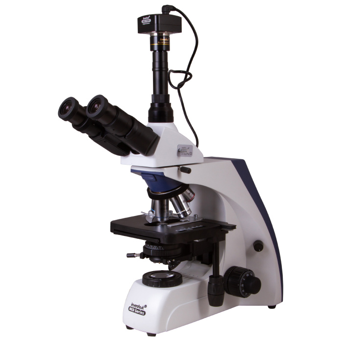
Levenhuk MED D35T Digital Trinocular Microscope
€1,569.95
Contattaci per una riservazione!
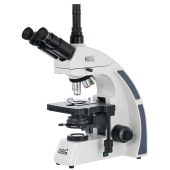
The Levenhuk MED D35T digital microscope for scientific research combines the functions of a classic biological model and a microscope for taking digital videos and photos. It is equipped with a 10 MP camera, connected to a computer, which allows real-time observation of an image on the screen. The microscope is ideal for a university department, a research center, and clinical or diagnostic laboratories.
| EAN/UPC | 5905555005041 |
|---|---|
| Manufacturer | LEVENHUK |
| Manufacturer Code | 74002 |
| Price | €1,569.95 |
Descrizione
The Levenhuk MED D35T digital microscope for scientific research combines the functions of a classic biological model and a microscope for capturing digital videos and photos. It is equipped with a 10 MP camera connected to a computer, allowing the observation of an image on the screen in real time. The microscope is ideal for a university department, a research center, and clinical or diagnostic laboratories.
Plan achromatic optics with infinity correction
The Levenhuk MED 35 series microscopes are equipped with an infinity-corrected optical system, used in professional and high-end microscopes. This system includes plan achromatic objectives and enables the transmission of clear, high-contrast images with an excellent level of flatness.
One of the most important features of the infinity-corrected optical system is that it allows the installation of additional parts in the optical path between the objective lens and the eyepiece. Additional parts include a polarizer for epi-fluorescence and filters. Overall, the modular design and ease of use make Levenhuk MED 35 microscopes optimal tools for various fields of microscopy, suitable for work in hematology, histology, microbiology labs, and much more.
The Levenhuk MED 35 series includes scientific microscopes for laboratory work
A trinocular head has a binocular viewing unit and a 30° inclined vertical tube for installing a digital camera. A beam splitter is present. The head rotates 360°, allowing convenient observation of the sample, especially if multiple researchers need to view it.
Optical capabilities
Experimental and research tasks are more practical and precise with the Levenhuk MED D35T microscope. The image is composed of 10x wide-field eyepieces with diopter adjustment and five plan achromatic objectives with various magnifications. The plan achromatic lenses flatten the field of view, reduce chromatic aberrations, and improve color rendition. Very detailed observations can be made with magnifications from 40x to 1000x. The 40x, 60x, and 100x objectives feature spring-loaded protective mounts. The 100x objectives are used for oil immersion observations. Sharpness is adjusted with coarse and fine focus knobs for macrometric and micrometric focusing.
Comfortable work with slides
The stage can be moved along two axes and is equipped with a mechanical graduated scale. Below the stage is a halogen light (30 W) with brightness adjustment. An Abbe condenser with an iris diaphragm is used to direct the light beam. The kit includes three optical filters. Additionally, Köhler illumination can be set up. The light is powered by AC power supply.
10 MP digital camera for capturing photos and videos
The 10 MP sensor of the digital camera (included in the kit) allows taking high-resolution photos. Additionally, it is possible to record videos without flicker at high frame rates.
The camera transmits the image to the screen of a connected computer. This makes laboratory work easier and more comfortable, as well as reduces strain on the eyes and shoulder joints, since there is no need to look through eyepieces. Special software (included on CD) allows easy processing of recorded images. The software enables modifying image size, brightness and contrast, exposure time, and white balance, calibrating the camera and objectives, as well as measuring samples and structures (various units of measurement available). The digital camera is powered and connected to the computer via USB cable.
Use of the Levenhuk MED D35T microscope in scientific settings ensures precise observations, allows application of the latest technologies, and enables long experiments in a practical way.
Features:
- Magnification: from 40x to 1000x
- Trinocular head with beam splitter and wide-field eyepieces
- Plan achromatic lenses with antifungal coating
- Halogen light with brightness adjustment
- Possibility to install Köhler illumination
- Powered via AC power supply
- 10 MP digital camera included
The kit includes:
- Microscope base with stand
- Rotatable trinocular head 360°
- Infinity-corrected plan achromatic objectives: 4x, 10x, 40xs, 60xs, 100xs (oil) with antifungal coating
- Wide-field eyepieces: WF10x/22 mm with antifungal coating (2 pieces)
- Abbe condenser N.A. 1.25 with iris diaphragm
- Filters: blue, green, yellow
- Bottle of immersion oil
- Fuse (2 pieces)
- Power cord for microscope
- Dust cover
- 10 MP digital camera
- Camera adapter
- Camera mount
- USB cable to connect and power the digital camera
- Driver and software CD
- User manual and lifetime warranty
Warning:
For information regarding the correct mains voltage, consult the specification table. Never attempt to connect a 110 V device to a 220 V outlet and vice versa without using a voltage converter. Note that mains voltage is 220–240 V in most European countries and 110 V in the United States and Canada.
Some examples of what can be observed under the microscope:


The Levenhuk MED D35T digital trinocular microscope is compatible with Levenhuk digital cameras (sold separately). Levenhuk cameras are installed on the optical tube in place of an eyepiece.
| Brand | LEVENHUK (USA) |
| Warranty, years | lifetime |
| EAN | 5905555005041 |
| Package size (LxWxH), cm | 62x36x25 |
| Shipping weight, kg | 10.92 |
| Type | digital, light/optical, biological |
| Head | trinocular |
| Optics material | optical glass with antifungal coating |
| Vertical tube | rotatable 360°, with beam splitter switch |
| Head inclination angle | 30° |
| Magnification, x | 40 — 1000 |
| Magnification range | from 800x to 1280x |
| Eyepiece tube diameter, mm | 30 mm (binocular head), 23.2 mm (third vertical tube) |
| Eyepieces | WF10x/22 mm, wide field with diopter adjustment (2 pcs.) |
| Objectives | infinity-corrected plan achromatic objectives: 4x, 10x, 40xs, 60xs, 100xs (oil) |
| Objective turret | for 5 objectives |
| Interpupillary distance, mm | 48 — 75 |
| Stage, mm | 180x160 |
| Stage movement range, mm | 80/50 |
| Stage features | double mechanical layer |
| Eyepiece diopter adjustment, diopters | ±5 |
| Condenser | Abbe condenser with N.A. 1.25 and iris diaphragm |
| Diaphragm | iris |
| Focus | coaxial, coarse (0.5 mm) and fine (0.002 mm), with rack and pinion |
| Body | metal |
| Illumination | halogen |
| Brightness adjustment | yes |
| Power supply | 100–240V |
| Light source type | 12 V/30 W, 85–230 V AC |
| Optical filters | blue, green, yellow |
| Extra components | collector, Köhler illumination |
| Megapixels | 10 |
| Sensor element | 1/2,3' |
| Pixel size, µm | 1.67х1.67 |
| Sensitivity, V/lux-sec@550 nm | 0.31 |
| Video recording | yes |
| Frame rate | 3.3@3584x2748 11@1792x1374 38@896x684 |
| Dynamic range, dB | 65.2 |
| Usage position | the third 23.2 mm eyepiece tube of the microscope |
| Image format | *.jpg, *.bmp, *.png, *.tif, etc. |
| Spectral range, nm | 380-650 |
| White balance | automatic/manual |
| Exposure control | 0.4–2000 μs |
| Software, driver | Levenhuk |
| Programmable options | image size, brightness, shutter speed |
| Output | USB 2.0 |
| System requirements | Windows 7/8/10 (32 and 64 bit), compatible with Mac OS 10.12 and Linux Ubuntu 14.04, Intel Core 2 processor or higher up to 2.8 GHz, at least 2 GB RAM, USB port, CD-ROM drive |
| Camera power supply | via USB cable |
| User level | expert users, professionals |
| Assembly and installation difficulty level | complicated |
| Video format | recording: *.wmv, *.avi, *.h264 (Windows 8 and later), *h265 (Windows 10 and later) |
| Application | laboratory/medical |
| Illumination position | lower |
| Research method | bright field |
| Set with case/carrying bag | dust cover |

Login and Registration Form