Customers Who Bought This Item Also Bought
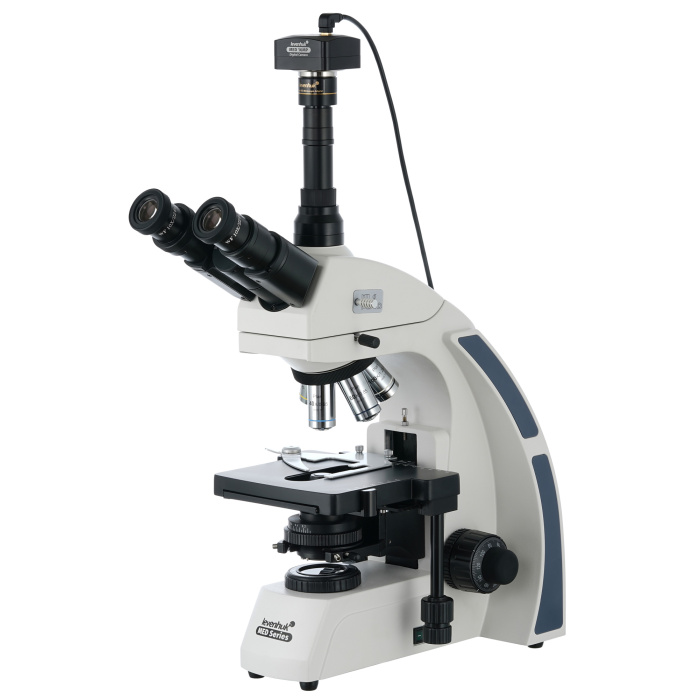
Levenhuk MED D40T Digital Trinocular Microscope
€1,729.95
Contattaci per una riservazione!
The Levenhuk MED D40T is a digital microscope with a trinocular head and a 16 MP camera included in the kit. The infinity-corrected planachromatic optical system produces a flat field, eliminates chromatic aberration, and allows observations with a high degree of detail. Köhler illumination can be used in the microscope, and oil immersion is available. The Levenhuk MED D40T is a perfect optical instrument for a professional who needs a microscope with extended functionality and high-quality optics.
| EAN/UPC | 5905555005140 |
|---|---|
| Manufacturer | LEVENHUK |
| Manufacturer Code | 74007 |
| Price | €1,729.95 |
Descrizione
Levenhuk MED D40T is a digital microscope with a trinocular head and a 16 MP camera included in the kit. The plan-achromatic infinity-corrected optical system produces a flat field, eliminates chromatic aberration, and allows observations with a high level of detail. Köhler illumination can be used with the microscope, and oil immersion is available. Levenhuk MED D40T is a perfect optical instrument for a professional who needs a microscope with extended functionality and high-quality optics.
Plan-achromatic optics with infinity correction
Microscope models in the Levenhuk MED 40 series come with an infinity-corrected optical system, used in professional and high-end microscopes. This system includes plan-achromatic objectives and enables the transmission of clear, high-contrast images with an excellent level of flatness.
One of the most important features of the infinity-corrected optical system is that it allows the installation of additional components in the optical path between the objective lens and the eyepiece. Additional components include a polarizer for epi-fluorescence and filters. Overall, the modular design and ease of use make Levenhuk MED 40 microscopes optimal tools for use in various fields of microscopy, for work in hematology, histology, microbiology laboratories, and much more.
Rotatable head with beam splitter
The trinocular head is standard for microscopes equipped with a digital camera in the kit. The binocular part is used for visual observations, and the camera is installed in a separate eyepiece tube. It is not necessary to interrupt the study to take a photo or record a video of samples. The visual module is slightly tilted at 30°, thus preventing neck muscle stiffness during long hours of observation. The head can be rotated 360°, and a beam splitter is present.
Optical capabilities
The kit includes optical accessories such as wide-field eyepieces with diopter adjustment and plan-achromatic objective lenses with various magnifications. The microscope’s total magnification ranges from 40x to 1000x. Three high magnification objectives have spring-loaded mounts. A 100x objective is intended for oil immersion. All optics are protected with antifungal coating.
16 MP digital camera for capturing photos and videos
A 16 MP digital camera allows taking high-resolution photos and recording videos. The resulting images can be used for professional work with micro-samples and for creating digital archives of studies. The camera connects to a PC via a USB cable (cable included in the kit) and transmits the image to the screen online. The camera transmits images to the screen of a connected computer. This makes laboratory work easier and more comfortable, while reducing eye and shoulder joint strain since it is unnecessary to observe through the eyepieces. Basic processing of recorded content can be performed using special software (CD included in the kit). The software allows adjusting image size, brightness and contrast, exposure time, and white balance, calibrating the camera and objective lenses, as well as measuring samples and structures (various units of measurement available).
Comfortable work with slides
Samples are placed under the objective using a mechanical stage. The stage can move in two directions. Coarse and fine focus are used to adjust sharpness. Intense LED light is positioned on the base. The illumination system also includes an Abbe condenser with an iris diaphragm. Köhler illumination is available.
The light is powered by an AC power supply. The brightness of the light is adjustable.
Features:
- Infinity-corrected plan-achromatic optics
- Trinocular head magnification range with beam splitter from 40x to 1000x
- Intense LED light with adjustable brightness
- Köhler illumination available
- Powered by AC adapter, brightness adjustable
- Includes 16 MP digital camera
The kit includes:
- Microscope base with stand
- 360° rotatable trinocular head
- Infinity-corrected plan-achromatic objective lenses: 4X, 10x, 40xs, 60xs, 100xs (oil) with antifungal coating
- Wide-field eyepieces: WF10x/22 mm with antifungal coating (2 pcs.)
- Abbe condenser N.A. 1.25 with iris diaphragm
- Filters: blue, green, yellow
- Immersion oil bottle
- Fuses (2 pcs.)
- Power cable for microscope
- Dust cover
- 16 MP digital camera
- Camera adapter
- Camera mount
- USB cable for connecting and charging the camera
- CD with software and drivers
- Lifetime user manual and warranty
Warning:
For information regarding correct mains voltage, refer to the specifications table. Never attempt to plug a 110 V device into a 220 V outlet and vice versa without using a voltage converter. Note that mains voltage is 220–240 V in most European countries and 110 V in the United States and Canada.
Some things that can be observed under the microscope:


. The Levenhuk MED D40T digital trinocular microscope is compatible with Levenhuk digital cameras (sold separately). Levenhuk cameras are installed on the optical tube, replacing an eyepiece.
| Brand | LEVENHUK (USA) |
| Warranty, years | lifetime |
| EAN | 5905555005140 |
| Package dimensions (LxWxH), cm | 50x44x28 |
| Shipping weight, kg | 10.34 |
| Type | digital, light/optical, biological |
| Head | trinocular |
| Optical material | optical glass with antifungal coating |
| Vertical tube | 360° rotatable, with beam splitter switch |
| Head tilt angle | 30° |
| Magnification, x | 40 — 1000 |
| Magnification range | from 800x to 1280x |
| Eyepiece tube diameter, mm | 30 mm (binocular head), 23.2 mm (third vertical tube) |
| Eyepieces | WF10x/22 mm, wide field with diopter adjustment (2 pcs.) |
| Objectives | infinity-corrected plan-achromatic objectives: 4x, 10x, 40xs, 60xs, 100xs (oil) |
| Objective turret | for 5 objectives |
| Interpupillary distance, mm | 48 — 75 |
| Stage, mm | 180x150 |
| Stage movement range, mm | 75/50 |
| Stage features | double-layer mechanical |
| Diopter adjustment of eyepiece, diopters | ±5 |
| Condenser | Abbe condenser with N.A. 1.25 and iris diaphragm |
| Diaphragm | iris |
| Focus | coaxial, coarse (0.5 mm) and fine (0.002 mm), rack and pinion |
| Body | metal |
| Illumination | LED |
| Brightness adjustment | yes |
| Power supply | 100–240V |
| Light source type | 5 W, 85–230 V AC |
| Optical filters | blue, green, yellow |
| Extra elements | collector, Köhler illumination |
| Megapixels | 16 |
| Sensor element | 1/2,33' |
| Pixel size, µm | 1.335x1.335 |
| Video recording | yes |
| Frame rate | 2@4632x3488 8@2320x1740 11@1536x1160 |
| Usage position | the third microscope eyepiece tube of 23.2 mm |
| Image format | *.jpg, *.bmp, *.png, *.tif, etc. |
| Spectral range, nm | 380-650 |
| White balance | automatic/manual |
| Exposure control | 0.2–2000 μs |
| Software, drivers | LevenhukLite |
| Programmable options | image size, brightness, shutter speed |
| Output | USB 2.0 |
| System requirements | Windows 7/8/10 (32 and 64 bit), compatible with Mac OS 10.12 and Linux Ubuntu 14.04, Intel Core 2 or higher processor up to 2.8 GHz, at least 2 GB RAM, USB port, CD-ROM drive |
| Camera power supply | via USB cable |
| User level | advanced users, professionals |
| Assembly and installation difficulty level | complicated |
| Video format | recording: *.wmv, *.avi, *.h264 (Windows 8 and later), *h265 (Windows 10 and later) |
| Application | laboratory/medical |
| Illumination position | lower |
| Research method | bright field |
| Digital camera included | yes |
| Maximum resolution | 4632x3488 |
Login and Registration Form