
Customers Who Bought This Item Also Bought
Levenhuk MED D10T Digital Trinocular Microscope
€829.95
Contattaci per una riservazione!
| EAN/UPC | 5905555004884 |
|---|---|
| Manufacturer | LEVENHUK |
| Manufacturer Code | 73986 |
| Price | €829.95 |
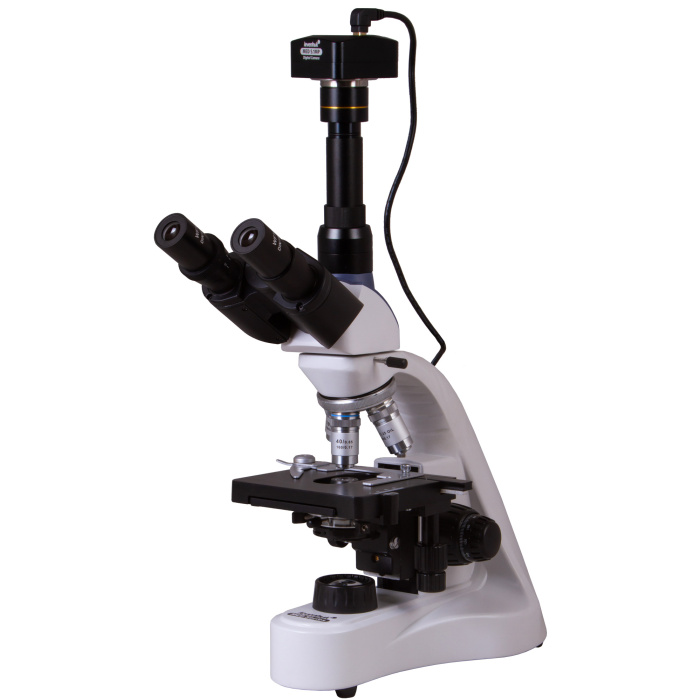
The Levenhuk MED D10T digital microscope is a classic trinocular model with a digital camera included in the kit. It is an excellent assistant for studying the microworld with magnifications ranging from 40x to 1,000x and for recording observations in photo or video mode. The microscope is suitable for holding lessons and seminars, conducting professional biological, clinical, and diagnostic research as well as research in various scientific fields. The microscope's achromatic optics are excellent for high-precision work with samples.
Reviews
Write Your Own Review
Descrizione
The Levenhuk MED D10T digital microscope is a classic trinocular model with a digital camera included in the kit. It is an excellent tool for studying the microworld with magnifications ranging from 40x to 1,000x and for recording observations in photo or video mode. The microscope is suitable for conducting lectures and seminars, carrying out professional biological, clinical, and diagnostic research, as well as research in various scientific fields. The microscope's achromatic optics are excellent for high-precision work with samples.
Tested rotating stage
The trinocular head consists of a binocular viewing part (intended for observations) and an eyepiece tube (for installing a digital camera). Due to this, it is possible to study a sample and simultaneously transmit an image to an external screen. This microscope makes it a perfect choice for group research when several people observe the same sample. The viewing part is inclined at 30°. It is comfortable to use during long observation sessions and also reduces strain on the neck muscles and shoulder girdle. Additionally, the head is rotatable 360°.
High-quality optics
The revolving nosepiece is designed for four objective lenses. The kit includes objective lenses of various magnifications — the 40x and 100x objective lenses are equipped with spring-loaded mounts to protect the optics from accidental damage. A 100x objective lens is suitable for oil immersion observations (a bottle of immersion oil is included in the kit). The wide-field eyepieces provide a good view and 10x magnification. All optical components of the microscope feature antifungal coating.
Compatible with digital cameras
The 5.1 MP digital camera is installed in the microscope’s eyepiece tube. The camera can operate in photo or video mode. It connects to a computer with pre-installed image processing software (a software CD and USB cable are included in the kit). The camera makes lab work easier and more convenient for researchers: there is no need to strain eyes by looking through eyepieces when everything is visible on a computer screen. This special software allows easy processing of photos and videos, setting shooting parameters, and saving data for later work. The software enables image size adjustments, brightness, contrast, and exposure time calibration of the camera and objective lenses, and also allows measuring samples and structures (various measurement units available). The high image quality allows usage for professional research.
Comfortable work with slides
The stage is movable along two axes and equipped with a mechanical scale. The microscope illumination system is located underneath. It consists of a powerful 5 W LED light and an Abbe condenser with an iris diaphragm and filter holder (three light filters are included). The light is powered by AC power supply. The illumination system also provides brightness adjustment. Coarse and fine focus adjustments are used to set image sharpness.
Features:
• Laboratory microscope with 40–1000x magnification
• 360° rotatable trinocular head, inclined at 30°
• Achromatic optics with antifungal coating
• 5 W LED transmitted light with brightness adjustment
• Powered by AC adapter
• 5.1 MP digital camera included in the kit
Kit includes:
• Microscope base with stand
• 360° rotatable trinocular head
• Achromatic objective lenses: 4x, 10x, 40xs, 100xs (oil) with antifungal coating
• Wide-field WF10x/18 mm eyepieces with antifungal coating (2 pcs.)
• Abbe condenser N.A. 1.25 with iris diaphragm and filter holder
• Filters: blue, green, yellow
• Bottle of immersion oil
• Fuse (2 pcs.)
• Microscope power cable
• Dust cover
• 5.1 MP digital camera
• Camera adapter
• USB cable for connecting and powering the camera
• Software CD and drivers
• User manual and lifetime warranty
Attention:
For correct mains voltage information, refer to the specifications table — never attempt to connect a 110 V device to a 220 V outlet or vice versa without using a converter. Bear in mind that the mains voltage is 220–240 V in most European countries and 110 V in the USA and Canada.
Some things you can observe with a microscope:


The Levenhuk MED D10T digital trinocular microscope is compatible with Levenhuk digital cameras (sold separately). Levenhuk cameras should be installed on the eyepiece tube, replacing an eyepiece.
| Brand | LEVENHUK (USA) |
| Warranty, years | lifetime |
| EAN | 5905555004884 |
| Package size (LxWxH), cm | 47.5x38.5x24 |
| Shipping weight, kg | 5.8 |
| Type | digital, light/optical, biological |
| Head | trinocular |
| Optics material | optical glass with antifungal coating |
| Vertical tube | 360° rotatable |
| Head tilt angle | 30° |
| Magnification, x | 40 — 1000 |
| Magnification range | from 800x to 1280x |
| Eyepiece tube diameter, mm | 23.2 |
| Eyepieces | WF10x/18 mm, wide field with diopter adjustment (2 pcs.) |
| Objectives | achromatic: 4x, 10x, 40xs, 100xs (oil immersion) |
| Revolving nosepiece | for 4 objectives |
| Interpupillary distance, mm | 48 — 75 |
| Stage dimensions, mm | 125х130 |
| Stage travel range, mm | 70/50 |
| Stage features | dual-layer mechanical |
| Eyepiece diopter adjustment, diopters | ±5 |
| Condenser | Abbe N.A. 1.25 with iris diaphragm and filter holder |
| Diaphragm | iris |
| Focus | coaxial, coarse (30 mm) and fine (0.002 mm) |
| Body | metal |
| Illumination | LED |
| Brightness control | yes |
| Power supply | 100–240V |
| Light source type | 5 W |
| Optical filters | blue, green, yellow |
| Megapixels | 5.1 |
| Sensor element | 1/2,5' |
| Pixel size, µm | 2.2х2.2 |
| Sensitivity, V/lux-sec@550 nm | 0.53 |
| Video recording | yes |
| Frame rate | 7@2592x1944 27@1280x960 90@640x480 |
| Dynamic range, dB | 66 |
| Usage position | the microscope's third 23.2 mm eyepiece tube |
| Image format | *.jpg, *.bmp, *.png, *.tif, etc. |
| Spectral range, nm | 380-650 |
| White balance | automatic/manual |
| Exposure control | 0.294–2000μs |
| Software, drivers | LevenhukLite |
| Programmable options | image size, brightness, shutter time |
| Output | USB 2.0 |
| System requirements | Windows 7/8/10 (32 and 64 bit), compatible with Mac OS 10.12 and Linux Ubuntu 14.04, Intel Core 2 processor or higher up to 2.8 GHz, at least 2 GB RAM, USB port, CD-ROM drive |
| User level | advanced users, professionals |
| Assembly and installation difficulty level | complicated |
| Video format | recording: *.wmv, *.avi, *.h264 (Windows 8 and later), *h265 (Windows 10 and later) |
| Application | laboratory/medical |
| Illumination position | lower |
| Research method | bright field |
| Digital camera included | yes |
| Set with case/carrying bag | dust cover |
| Maximum resolution | 2048x1536 |

Login and Registration Form